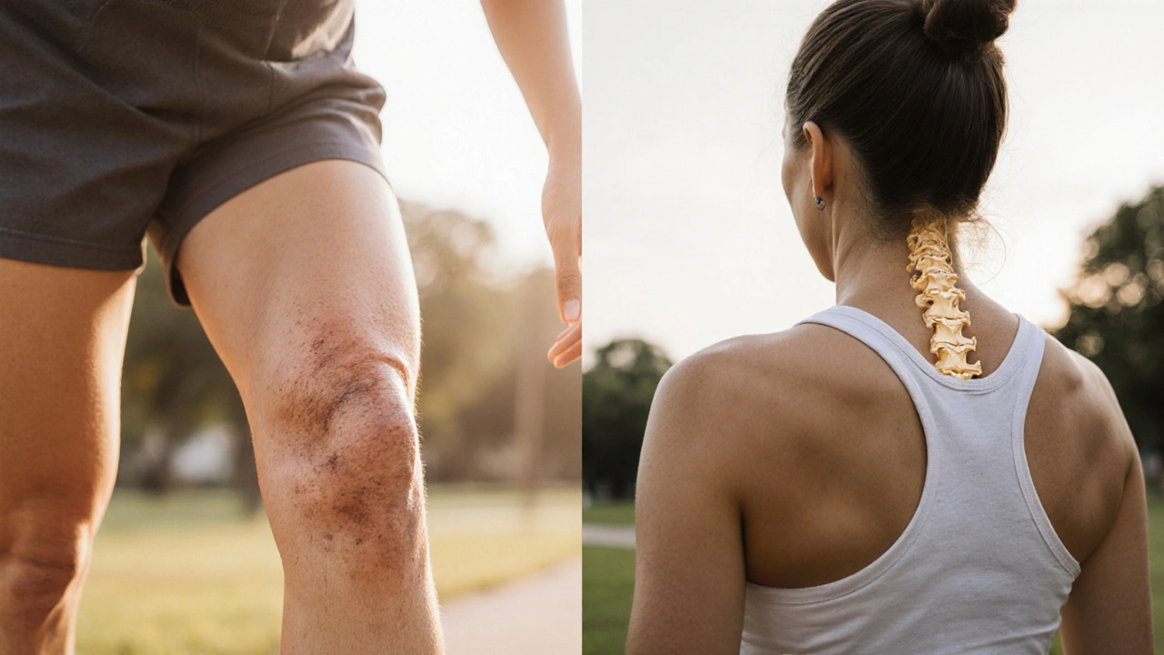
Osteoarthritis is a chronic joint disease that primarily damages cartilage and the underlying bone, leading to pain, stiffness, and reduced mobility. It affects up to 10% of adults over 60 and is the most common form of arthritis worldwide.
Many people assume that having a joint problem protects the bones, but recent research shows a surprising overlap with Osteoporosis, a condition where bone mineral density drops enough to raise fracture risk. Osteoporosis affects roughly one in three women and one in five men over 50. Understanding how these two disorders interact helps clinicians tailor treatment and gives patients clearer guidance on lifestyle choices.
Key Takeaways
- Both osteoarthritis and osteoporosis share age‑related, hormonal, and lifestyle risk factors.
- Joint degeneration can accelerate bone loss by altering loading patterns on subchondral bone.
- Conversely, low bone density can worsen joint pain and increase the chance of osteoarthritis‑related fractures.
- Integrated screening-using bone density scans and joint imaging-detects the overlap early.
- Exercise, nutrition, and medication choices can address both conditions simultaneously.
What Is Osteoarthritis?
Osteoarthritis (OA) begins with cartilage degradation. Articular cartilage loses its proteoglycan matrix, becomes thinner, and eventually cracks. The exposed bone surface triggers subchondral bone remodeling, osteophyte formation, and synovial inflammation. Commonly affected joints include knees, hips, hands, and the spine.
Risk factors are multifactorial:
- Age - prevalence rises sharply after 50.
- Genetics - certain collagen‑type genes increase susceptibility.
- Obesity - excess weight adds mechanical stress and inflammatory cytokines.
- Joint injury - previous trauma accelerates cartilage wear.
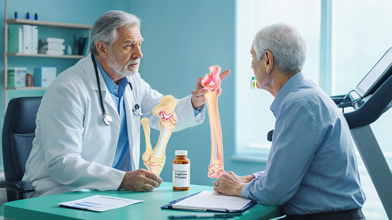
What Is Osteoporosis?
Osteoporosis (OP) is defined by a drop in bone density. A dual‑energy X‑ray absorptiometry (DEXA) T‑score of -2.5 or lower signals the disease. The skeleton becomes porous, making even minor falls capable of causing fractures, especially in the hip, vertebrae, and wrist.
Key contributors include:
- Hormonal changes - estrogen loss in post‑menopausal women is a major driver.
- Insufficient calcium and vitamin D. Vitamin D facilitates calcium absorption and bone mineralization..
- Physical inactivity - weight‑bearing exercise stimulates bone formation.
- Smoking and excessive alcohol - both impair osteoblast function.
Shared Risk Factors Linking OA and OP
Although OA and OP affect different tissues, they converge on several systemic and lifestyle factors.
- Inflammation. Cytokines such as IL‑6 and TNF‑α promote cartilage breakdown and increase osteoclast activity.
- Age‑related hormonal shifts - reduced estrogen and growth hormone affect both cartilage health and bone remodeling.
- Vitamin D deficiency - lowers calcium absorption and may impair muscle function, leading to altered gait that stresses joints.
- Low calcium intake. Calcium is essential for both cartilage matrix synthesis and bone mineral density..
- Physical inactivity - lack of weight‑bearing activity diminishes bone loading and reduces synovial fluid circulation, aggravating both conditions.
How Joint Degeneration Affects Bone Density
When cartilage erodes, the underlying subchondral bone bears more load. This altered stress triggers two processes:
- Subchondral sclerosis. Bone becomes denser but more brittle, paradoxically lowering overall bone quality.
- Reduced mobility - painful joints limit walking or resistance training, decreasing the mechanical stimulus needed for bone maintenance.
Studies from Australian bone health registries show that knee OA patients who limit activity for more than six months can lose up to 2% of lumbar spine BMD per year, a rate comparable to untreated osteoporosis.
How Low Bone Density Impacts Joint Health
Weak bones change the way forces are transmitted through a joint. A fragile femoral head, for example, can micro‑fracture under normal walking loads, leading to altered joint alignment and increased cartilage wear.
Furthermore, vertebral compression fractures cause postural changes that increase load on hip and knee joints, accelerating osteoarthritic changes in those areas.
Clinical Implications: Diagnosis and Integrated Management
Given the overlap, clinicians should adopt a dual‑screening approach:
- For patients with symptomatic OA, add a DEXA scan if they are over 55, have a history of fractures, or present with low BMI.
- For osteoporosis patients reporting joint pain, request plain radiographs or MRI to assess cartilage status.
Management can often target both diseases:
- Exercise: Weight‑bearing and resistance training improve BMD while enhancing joint stability. Taichi and low‑impact aerobics are OA‑friendly.
- Nutrition: Adequate calcium (1,200mg/day for adults over 50) and vitamin D (800-1,000IU/day) support bone and cartilage health.
- Medication synergy: Bisphosphonates reduce bone resorption and may modestly slow subchondral bone changes in OA. NSAIDs control OA pain but should be used cautiously in osteoporosis patients with gastrointestinal risk.
- Fall prevention: Home safety assessments, balance training, and vision checks lower fracture risk for those with compromised joints.
Comparison Table: Osteoarthritis vs Osteoporosis
| Attribute | Osteoarthritis | Osteoporosis |
|---|---|---|
| Primary tissue affected | Articular cartilage & subchondral bone | Trabecular and cortical bone |
| Typical diagnostic test | Radiographs, MRI, clinical grading (Kellgren‑Lawrence) | DEXA scan (T‑score ≤ -2.5) |
| Common age of onset | 50‑70 years | 55‑75 years (earlier in women post‑menopause) |
| Major risk factors | Obesity, joint injury, genetics | Estrogen deficiency, low calcium, sedentary lifestyle |
| Key symptom | Joint pain, stiffness, reduced range of motion | Silent until fracture; back pain from vertebral collapse |
| Impact on bone density | Can cause localized sclerosis yet overall BMD may drop due to inactivity | Systemic loss of BMD across the skeleton |
Related Concepts and Next Steps
Understanding the link between OA and OP opens doors to broader topics such as metabolic bone disease, chronic pain management, and geriatric orthopedics. Readers interested in the hormonal side can explore "Estrogen’s role in bone remodeling". Those focused on lifestyle can dive into "Weight‑bearing exercise protocols for seniors". Finally, a deeper look at "Inflammatory cytokines in musculoskeletal aging" rounds out the knowledge cluster.
Frequently Asked Questions
Can having osteoarthritis protect me from osteoporosis?
No. While OA does increase bone formation in the affected joint, overall mobility often drops, which can accelerate systemic bone loss. In fact, many OA patients develop osteoporosis later if they stop exercising.
Should I get a DEXA scan if I have knee pain?
It’s advisable if you’re over 55, have a family history of fractures, or have low body weight. A DEXA scan can detect early osteoporosis, allowing you to start preventive treatment before a fracture occurs.
Do osteoporosis medications help osteoarthritis?
Bisphosphonates primarily target bone resorption, but some studies suggest they may slow subchondral bone changes that aggravate OA. They don’t replace OA‑specific treatments like physiotherapy or NSAIDs.
What type of exercise is safest for both conditions?
Low‑impact, weight‑bearing activities such as walking, elliptical training, and resistance band work are ideal. They reinforce bone density while sparing joints from high‑impact stress.
How does vitamin D influence both OA and OP?
Vitamin D promotes calcium absorption, essential for bone mineralization, and also modulates inflammatory pathways in cartilage. Deficiency is linked to higher pain scores in OA and lower BMD in OP.
 Liver Disease and Drug Metabolism: How Reduced Clearance Affects Medication Safety
 Antabuse Online Prescription: A Comprehensive Guide
 How to Audit Your Medication Bag Before Leaving the Pharmacy: A Simple 7-Step Safety Check
 Terramycin vs Alternative Antibiotics: Tetracycline Comparison Guide
G.Pritiranjan Das
September 27, 2025 AT 17:33Stay active, keep those joints moving and your bones will thank you.
Karen Misakyan
October 1, 2025 AT 05:33One must acknowledge that the reciprocal influence between osteoarthritis and osteoporosis extends beyond mere coincidental comorbidity; it reflects a systemic alteration of biomechanical and metabolic pathways. The degradation of articular cartilage precipitates compensatory subchondral bone remodeling, which in turn modulates overall skeletal density. Moreover, inflammatory mediators such as interleukin‑6 and tumor necrosis factor‑α serve as common denominators that exacerbate both conditions. Consequently, clinicians are urged to adopt an integrated diagnostic algorithm that incorporates both radiographic joint assessment and densitometric bone evaluation. In practice, this approach facilitates early intervention, thereby mitigating the cascade of functional decline associated with these disorders.
vijay sainath
October 4, 2025 AT 17:33Alright, enough of the high‑falutin jargon – the real issue is that most patients just end up sitting on the couch because the pain meds make them drowsy, and then their bones get weaker faster than you can say “osteoporosis.” The article glosses over this simple cause‑and‑effect chain.
Daisy canales
October 8, 2025 AT 05:33Wow, groundbreaking insight there. Who would’ve thought that moving less makes you weaker?
Mason Grandusky
October 11, 2025 AT 17:33Man, reading about the OA‑OP crossover really lights a fire under me! Imagine crushing those myths with a combo of kettlebell swings and calcium‑rich smoothies – that’s the sweet spot where bone gets denser and joints stay supple. I swear, when you blend science with a dash of hype, the whole picture just clicks. Let’s keep the convo rolling, folks, because knowledge is the ultimate rep!
Emily Stangel
October 15, 2025 AT 05:33The interplay between osteoarthritis and osteoporosis is a classic illustration of how the skeletal system cannot be compartmentalized into isolated pathologies. When cartilage deteriorates, the subchondral bone is subjected to altered mechanical loads that may foster localized sclerosis while simultaneously reducing systemic bone turnover due to decreased activity. Conversely, a porous skeleton fails to provide the necessary support for joint congruity, precipitating micro‑fractures that disturb the articular surface. From a clinical perspective, this bidirectional relationship necessitates a paradigm shift toward integrated screening rather than siloed investigations. A patient presenting with knee pain over the age of 55 should be evaluated not only with radiographs but also with a DEXA scan to uncover occult osteoporosis. Similarly, an individual with a recent vertebral fracture who complains of hip discomfort warrants an MRI to rule out early osteoarthritic changes. Research consistently demonstrates that physical inactivity, often a consequence of joint pain, accelerates bone mineral density loss at a rate comparable to untreated osteoporosis. Weight‑bearing exercises such as brisk walking, resistance band work, and low‑impact aerobics simultaneously stimulate osteoblastic activity and improve joint lubrication. Nutritional adequacy, particularly calcium intake of 1,200 mg per day and vitamin D levels above 30 ng/mL, underpins both bone mineralization and cartilage matrix synthesis. Pharmacologically, bisphosphonates may offer a modest protective effect on subchondral bone remodeling, though they do not replace the need for analgesic and physiotherapeutic strategies specific to osteoarthritis. NSAIDs, while effective for pain control, must be prescribed judiciously in patients with documented gastrointestinal risk, a concern heightened in the elderly osteoporotic population. Fall prevention measures, including home safety assessments, balance training, and regular vision checks, constitute a non‑pharmacologic cornerstone in reducing fracture incidence. It is also prudent to monitor hormonal status, as estrogen deficiency amplifies both cartilage degradation and bone resorption. Emerging biomarkers, such as serum CTX and cartilage oligomeric matrix protein, hold promise for early detection of the osteoarthritis‑osteoporosis nexus. In summary, a holistic approach that intertwines musculoskeletal assessment, lifestyle modification, and targeted therapy offers the best prospect for preserving function and quality of life.
Suzi Dronzek
October 18, 2025 AT 17:33While the comprehensive overview is commendable, one must not lose sight of the ethical imperative to ensure equitable access to both DEXA scanning and physical therapy. It is disheartening that socioeconomic disparities continue to dictate who receives proper musculoskeletal care. The discourse should therefore emphasize policy reforms alongside clinical recommendations.
Aakash Jadhav
October 22, 2025 AT 05:33Listen up, comrades! The silent war between cartilage and calcium is raging within our bodies, and we are the unsuspecting soldiers. If we ignore the signs, the betrayal of our own skeleton will be our ultimate tragedy.
Amanda Seech
October 25, 2025 AT 17:33Totally get what u're sayin, Aakash. Yeah the joint pain can really mess up your mood and make you feel like u’re stuck in a rut.
Lisa Collie
October 29, 2025 AT 04:33Honestly, I find the entire premise of linking two distinct pathologies somewhat reductive; the literature often overstates correlation where mere coincidence resides.
Anthony Palmowski
November 1, 2025 AT 16:33Whoa!!! You really think the evidence is that flimsy??? I mean, come on, the studies are robust, statistically significant, and peer‑reviewed!!!
Jillian Rooney
November 5, 2025 AT 04:33America needs to stand up for its seniors and protect them from bone loss.
Rex Peterson
November 8, 2025 AT 16:33The dialectic between degenerative joint disease and systemic bone demineralization invites a metaphysical inquiry into the unity of the corporeal vessel. If the musculoskeletal system is conceived as an integrated whole, then fragmentation of one component inevitably perturbs the equilibrium of the entire organism.
Candace Jones
November 12, 2025 AT 04:33Great synthesis, Rex. Adding a note about balanced nutrition could further empower readers to take proactive steps.
Robert Ortega
November 15, 2025 AT 16:33Both conditions share modifiable risk factors, so public health initiatives targeting lifestyle could yield dual benefits.
Elizabeth Nisbet
November 19, 2025 AT 04:33Exactly, Robert! Small changes like taking the stairs or adding a daily glass of milk can make a big difference.
Sydney Tammarine
November 22, 2025 AT 16:33Behold! The saga of bone and cartilage unfurls before our very eyes, and we, mere mortals, must rise to the challenge 🌟.
josue rosa
November 26, 2025 AT 04:33In the context of mechanotransduction, alterations in subchondral microarchitecture precipitate a cascade of osteoclastic signaling pathways, thereby amplifying the catabolic milieu that underlies osteoporotic phenotypes. This bidirectional feedback loop underscores the necessity for interdisciplinary therapeutic regimens integrating rheumatologic pharmacodynamics with orthopedic biomechanics.
Shawn Simms
November 29, 2025 AT 16:33While the article is informative, it contains several instances of misplaced modifiers and inconsistent tense usage that could be corrected for greater clarity.